• There are no suggestions because the search field is empty.

Boost productivity with AI-assisted image analysis

Aiforia equips pathologists and scientists in clinical, pre-clinical, and academic labs with powerful deep learning AI and cloud-based technology. Aiforia’s solutions strive to increase the speed, accuracy, and consistency of analyzing large and complex medical images, especially in pathology. 

Request a demo
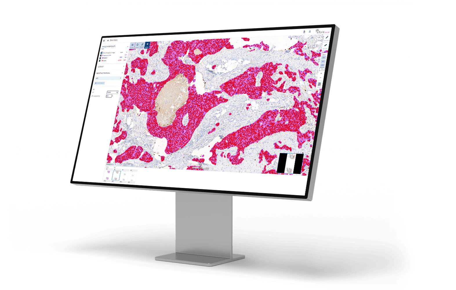
ER_monitor_right angle

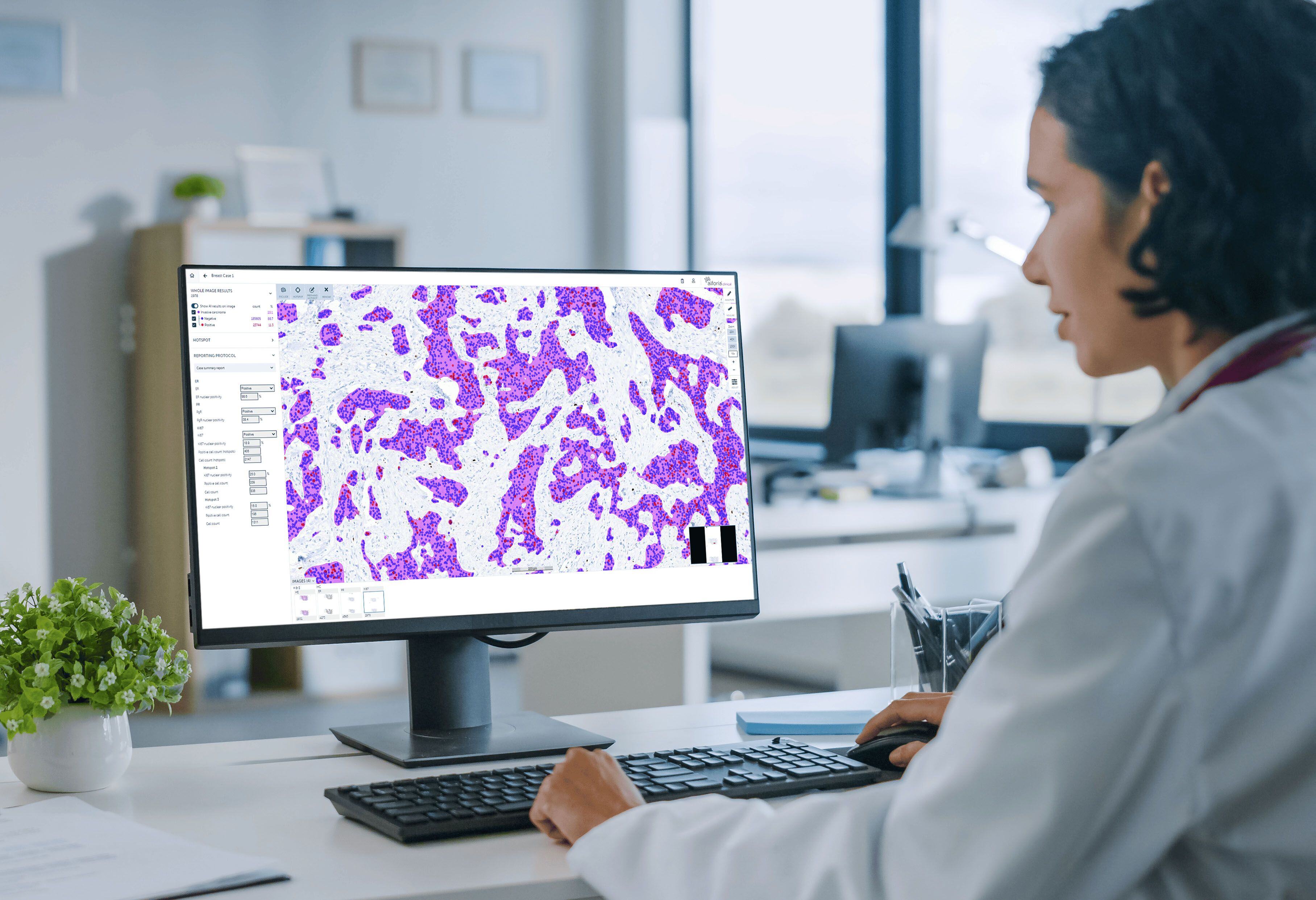
Clinical stock image_Ki67

Clinical solutions

For pathology labs looking to increase productivity and improve diagnostic accuracy, Aiforia's clinical solutions offer AI-supported image-based diagnostics with intelligent visualization, assisted screening, and reporting tools in one cloud-based platform. Clinical suites – Breast Cancer, Lung Cancer, Prostate Cancer, and Colorectal Cancer – consist of AI models and matching Aiforia® Clinical Suite Viewer*.

*Only certain Aiforia® Clinical AI models and the Aiforia® Clinical Suite Viewer are CE-IVD marked for diagnostic use in EU and EEA countries; see here for the full list: www.aiforia.com/aiforia-clinical-solutions. In all other countries, the use is limited to Research Use Only, not for use in diagnostic procedures.

Learn more
AI development tool

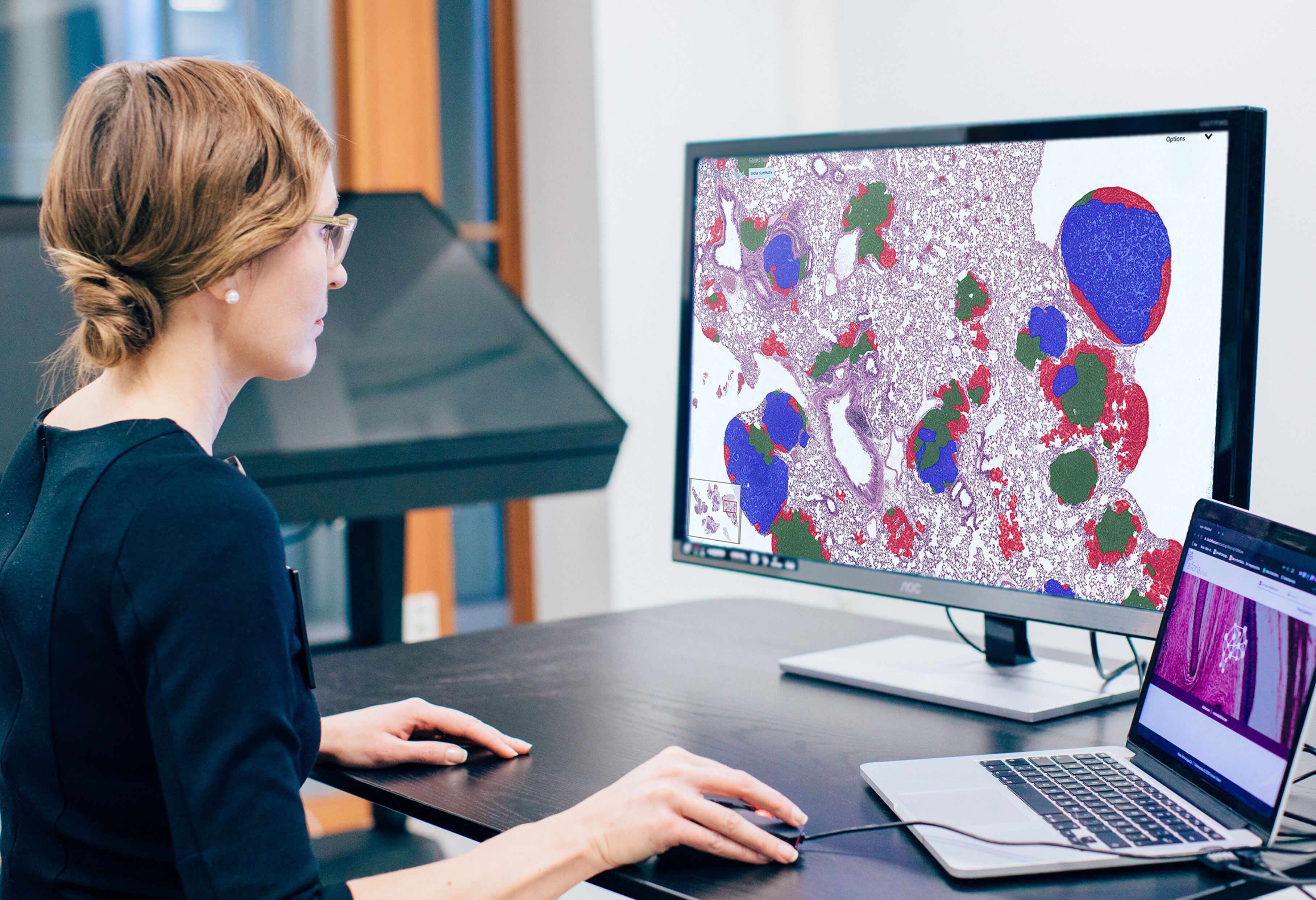
AI development tool

Aiforia®️ Create is the most versatile tool for developing, customizing, and validating deep learning AI models for histological features and patterns in image analysis. Its cloud-based, collaborative working environment allows multiple users to work together in real time, anywhere in the world. Praised for its intuitive user interface, it allows users a fast start, even without any prior AI experience. 

Learn more
Research solutions

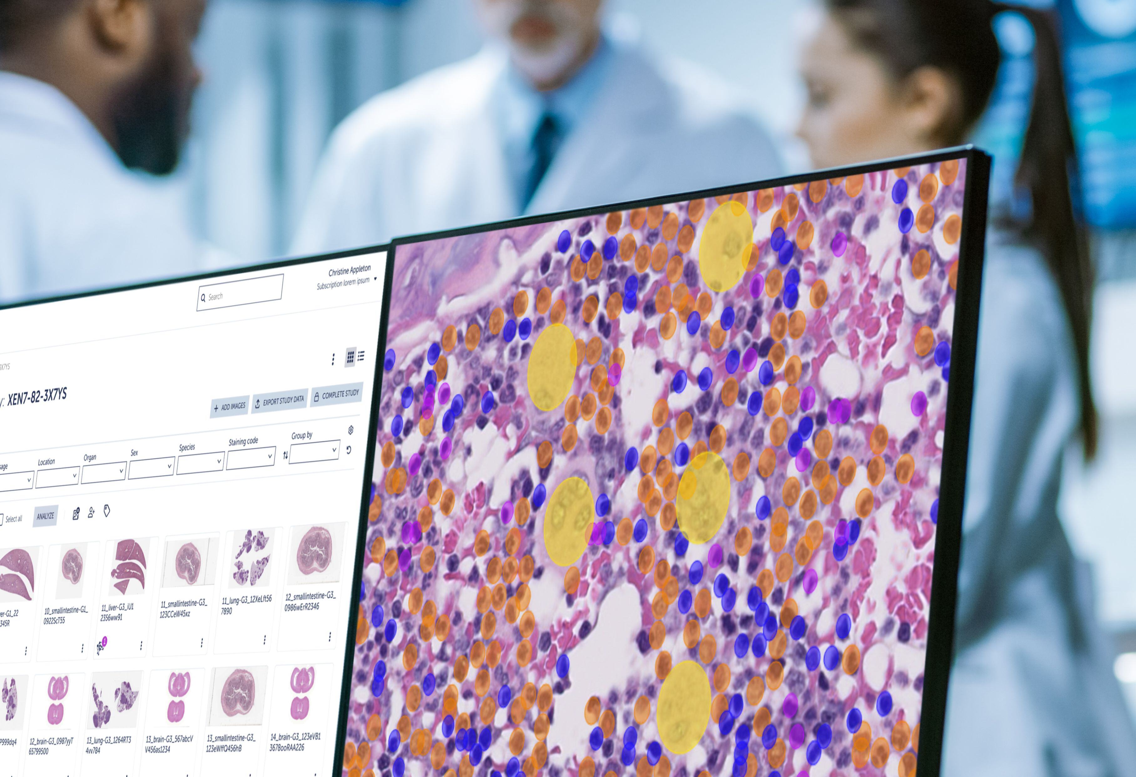
Research solutions

For pharma, biotech, CROs, and academic researchers, Aiforia's research solutions offer cloud-based pathology image analysis applications that cater to all research areas. Empowered by robust AI capabilities, pathologists and scientists can make the most of a digitized workflow and fully harness their expertise by automating repetitive tasks and increasing the speed and accuracy of study reviews.

Learn more
This is what our customers have to say

"I have used Aiforia for the past couple of years for two research projects with clinical utility. The platform is nicely done, with a myriad of options to analyze and evaluate results. The support staff have been wonderful, and they worked diligently with us every step of the way to accomplish our goals; I honestly have never seen this level of outstanding support."

Shadi Qasem, MD, MBA, Chief of Pathology at Baptist Health


"Our lab has found Aiforia to be an enormous asset to our research. Initially, we used it as a fast and accurate replacement for stereology and now we are pushing into new boundaries of image analysis. During the development of our first models, we received excellent support in understanding computer vision and learning strategies for training in object detection. The platform is well-designed and intuitive, making the learning curve relatively short. The ability to customize models to our various research interests is invaluable in part because the iterative process shows us more and more about our source images. It’s an excellent tool." 

Lucy Greene, Postgraduate Associate (Neuroscience), Yale University School of Medicine


"The Aiforia platform has empowered our pathology trainees to pursue exciting AI projects. The platform is built for pathologists and requires only basic understanding of AI principles. The Aiforia staff provides excellent training and support. I would recommend it to anyone without hesitation."

Lee Cooper, Director, Computational Pathology at Northwestern University

Why Aiforia?

ic_our_ai-1_pink

Future-proof

Together with our customers, we continue to develop new groundbreaking AI models, with thousands of models already developed for research use. We’re committed to providing cutting-edge deep learning AI models designed to meet the evolving needs of our customers. As pathology image analysis becomes increasingly complex, our advanced software will ensure you stay at the forefront of technology.

ic_discover_pink

Transparent

Aiforia commits to keeping our AI algorithms transparent and understandable. You remain in control, see how analyses are conducted, and participate in the training process. AI models serve as first-read support while the expert verifies the results. We offer our customers fully quantifiable data and easily adjustable results.

ic_wellbeing_pink

Easy to use

Aiforia’s image analysis software requires no data scientists or coding expertise. Our team of experts integrates the solutions into existing IT infrastructures. We acknowledge that every lab is different, so we optimize each model for each lab’s needs and monitor their performance to deliver continuous improvement. Aiforia’s software allows customers to confidently scale up their operations while paying only for what they need.

ic_secure_pink

Secure

We take data privacy and security very seriously. We hold several globally relevant quality and security certifications so our customers can be sure their sample and patient data is safe.* Customers determine what information is stored in the cloud and can opt for a private version.
 

*Our image analysis solutions are developed and maintained securely per ISO 27001 and SOC 2 Type II security standards, and ISO 13485 quality standards. In addition, we strictly adhere to GDPR requirements and all applicable HIPAA policies and procedures.


Request a demo

Discover the power of AI for image analysis

Learn how to enhance your image analysis work in diagnostic pathology, preclinical studies, and medical research. The demo will be tailored to your interests.

The demo will help you understand:

  • How AI-assisted image analysis can increase efficiency, precision, and consistency in the pathology workflow.
  • The limitless possibilities Aiforia® Platform offers – both for research and clinical diagnostics – and the suitable use cases for your needs.

We can either demonstrate Aiforia’s image analysis solutions on your images or any of our application examples (e.g., neuron quantification, automated tumor grading, NASH analysis).

Fill in the form, and one of our experts will contact you shortly.