• There are no suggestions because the search field is empty.
Aiforia® Breast Cancer Suite

AI for breast cancer diagnostics

Transform your pathology lab with Aiforia® Breast Cancer Suite – enhancing precision, speed, and diagnostic confidence with cutting-edge AI technology.

Aiforia® Breast Cancer Suite consists of AI models with an optimized interactive user interface that supports pathologists in the histological grading of breast cancer and in the assessment of breast cancer IHC markers.

Only certain Aiforia® Clinical AI models and the Aiforia® Clinical Suite Viewer are CE-IVD marked for diagnostic use in EU and EEA countries; see details under each AI model.

Discuss with a pathologist
Breast cancer suite

Enhance your pathology workflow with Aiforia

Aiforia® Breast Cancer Suite offers a fully digital cockpit for breast cancer diagnostics, supporting the entire pathology workflow from sample analysis to diagnosis.


Breast Cancer workflow

Breast Cancer Suite: AI models

Aiforia® Breast Cancer Grading
Aiforia® Breast Cancer ER
Aiforia® Breast Cancer PR
Aiforia® Breast Cancer HER2
Aiforia® Breast Cancer Ki67
Aiforia® Lymph Node Metastasis
breast-cancer-grading_image 1

The AI model supports the histological grading of breast cancer in whole slide images (WSI) of H&E stained tissue biopsies. It detects invasive carcinoma and ductal carcinoma in situ (DCIS) and scores the grading components mitotic count, tubulus formation, and nuclear pleomorphism. It has been designed based on the Nottingham Grading System. 


Aiforia® Breast Cancer Grading is currently for Research Use Only (RUO) and for Performance Studies Only (PSO) in all market areas. 

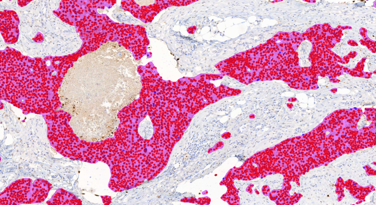
Breast ER with AI masks

The AI model supports the detection of invasive carcinoma and scoring of ER positive and negative tumor cells from whole slide images (WSI) or from selected image areas of cancerous breast tissue.

Clinical Validation
  • Showed excellent correlation with visual diagnosis
  • Saved time (up to 47% faster)


CE-IVD marked for diagnostic use in EU and EEA countries, and for Research Use Only (RUO) and Performance Studies Only (PSO) in all other market areas

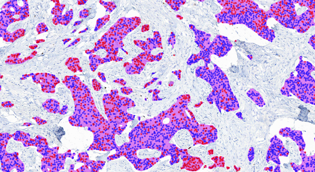
Breast PR with AI masks

The AI model supports the detection of invasive carcinoma and scoring of PR positive and negative tumor cells from whole slide images (WSI) or from selected image areas of cancerous breast tissue.

Clinical Validation
  • Showed excellent correlation with visual diagnosis
  • Saved time (up to 16% faster)


CE-IVD marked for diagnostic use in EU and EEA countries, and for Research Use Only (RUO) and Performance Studies Only (PSO) in all other market areas

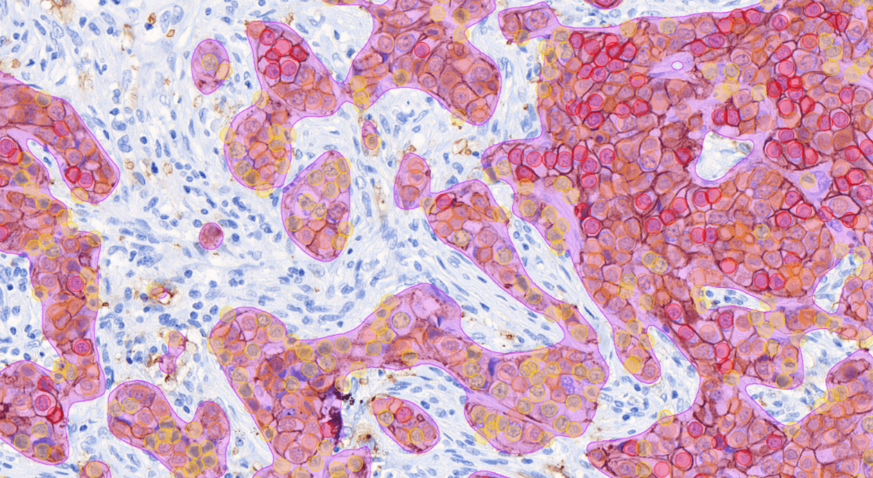
BrCa_HER2_10x_cellmask2-1

The Al model supports the detection of invasive carcinoma and scoring of HER2 positivity and intensity in tumor cells from whole slide images
(WSI) of breast tissue. The Al model has been designed according to CAP recommendations for HER2 analysis.

Clinical Validation
  • Showed excellent correlation with visual diagnosis
  • Improved diagnostic concordance by increasing pathologist agreement
  • Saved time (up to 16% faster)


Aiforia® Breast Cancer HER2 is currently for Research Use Only (RUO) and for Performance Studies Only (PSO) in all market areas. 

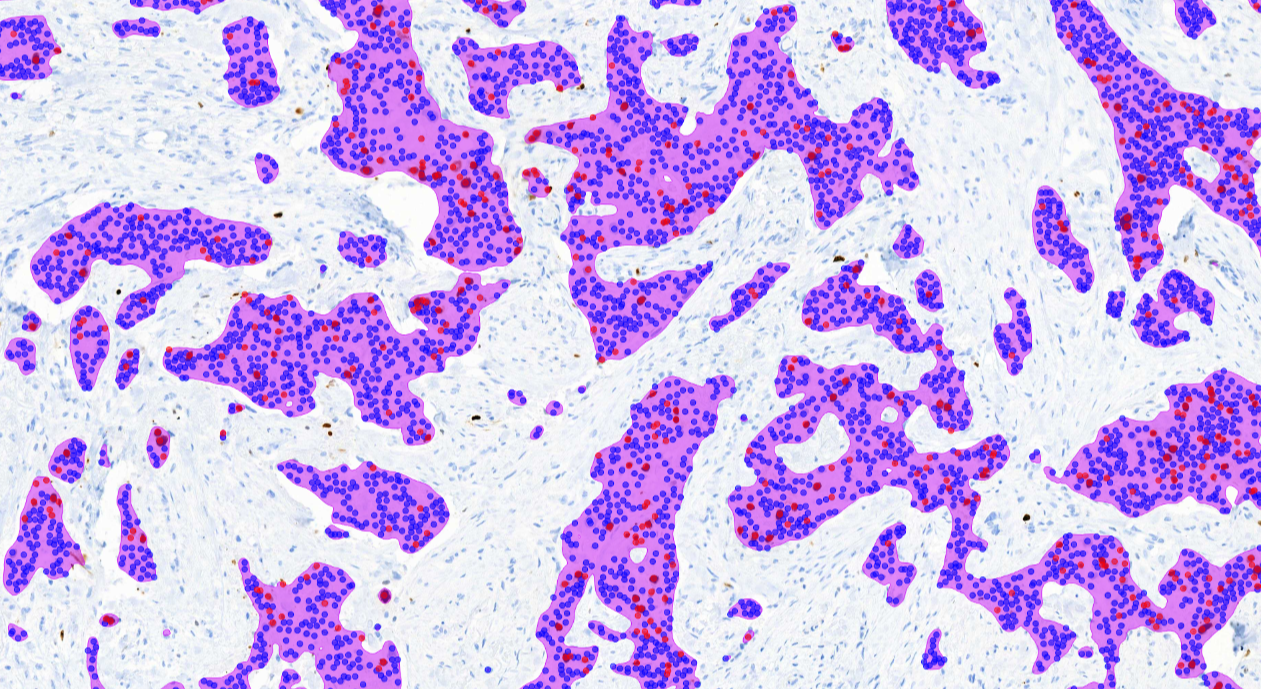
Breast Ki67 with AI masks

The AI model supports the detection of invasive carcinoma and scoring of Ki67 positive and negative tumor cells from whole slide images (WSI) or from selected image areas of cancerous breast tissue.

Clinical Validation
  • Showed excellent correlation with visual diagnosis
  • Improved diagnostic concordance by increasing pathologist agreement
  • Saved time in the whole slide image scoring (up to 54% faster)

CE-IVD marked for diagnostic use in EU and EEA countries, and for Research Use Only (RUO) and Performance Studies Only (PSO) in all other market areas

aiforia-lymph-node-metastasis-breast

The AI model supports the detection and quantification of metastases of breast cancer, melanoma, and colorectal cancer in lymph nodes from whole slide images.

Aiforia® Lymph Node Metastasis is currently for Research Use Only (RUO) and for Performance Studies Only (PSO) in all market areas. 

Breast Cancer Suite: AI models

Aiforia® Breast Cancer Grading

breast-cancer-grading_image 1

The AI model supports the histological grading of breast cancer in whole slide images (WSI) of H&E stained tissue biopsies. It detects invasive carcinoma and ductal carcinoma in situ (DCIS) and scores the grading components mitotic count, tubulus formation, and nuclear pleomorphism. It has been designed based on the Nottingham Grading System. 


Aiforia® Breast Cancer Grading is currently for Research Use Only (RUO) and for Performance Studies Only (PSO) in all market areas. 

Aiforia® Breast Cancer ER

Breast ER with AI masks

The AI model supports the detection of invasive carcinoma and scoring of ER positive and negative tumor cells from whole slide images (WSI) or from selected image areas of cancerous breast tissue.

Clinical Validation
  • Showed excellent correlation with visual diagnosis
  • Saved time (up to 47% faster)


CE-IVD marked for diagnostic use in EU and EEA countries, and for Research Use Only (RUO) and Performance Studies Only (PSO) in all other market areas

Aiforia® Breast Cancer PR

Breast PR with AI masks

The AI model supports the detection of invasive carcinoma and scoring of PR positive and negative tumor cells from whole slide images (WSI) or from selected image areas of cancerous breast tissue.

Clinical Validation
  • Showed excellent correlation with visual diagnosis
  • Saved time (up to 16% faster)


CE-IVD marked for diagnostic use in EU and EEA countries, and for Research Use Only (RUO) and Performance Studies Only (PSO) in all other market areas

Aiforia® Breast Cancer HER2

BrCa_HER2_10x_cellmask2-1

The Al model supports the detection of invasive carcinoma and scoring of HER2 positivity and intensity in tumor cells from whole slide images
(WSI) of breast tissue. The Al model has been designed according to CAP recommendations for HER2 analysis.

Clinical Validation
  • Showed excellent correlation with visual diagnosis
  • Improved diagnostic concordance by increasing pathologist agreement
  • Saved time (up to 16% faster)


Aiforia® Breast Cancer HER2 is currently for Research Use Only (RUO) and for Performance Studies Only (PSO) in all market areas. 

Aiforia® Breast Cancer Ki67

Breast Ki67 with AI masks

The AI model supports the detection of invasive carcinoma and scoring of Ki67 positive and negative tumor cells from whole slide images (WSI) or from selected image areas of cancerous breast tissue.

Clinical Validation
  • Showed excellent correlation with visual diagnosis
  • Improved diagnostic concordance by increasing pathologist agreement
  • Saved time in the whole slide image scoring (up to 54% faster)

CE-IVD marked for diagnostic use in EU and EEA countries, and for Research Use Only (RUO) and Performance Studies Only (PSO) in all other market areas

Aiforia® Lymph Node Metastasis

aiforia-lymph-node-metastasis-breast

The AI model supports the detection and quantification of metastases of breast cancer, melanoma, and colorectal cancer in lymph nodes from whole slide images.

Aiforia® Lymph Node Metastasis is currently for Research Use Only (RUO) and for Performance Studies Only (PSO) in all market areas. 

Why Aiforia?

Products_Clinical

The most comprehensive AI toolkit

Aiforia® Breast Cancer Suite offers advanced tools for histological grading and biomarker detection, ensuring precision and efficiency.

Industry_CRO

Transparent and adjustable results

Review accurate pixel-level results at the highest resolution. The suite improves repeatability and traceability while giving you full control of the results. 

ic_speed_up_pink

High lifetime value

Designed for longevity, Aiforia’s AI models evolve with your lab’s needs, ensuring consistent accuracy and flexibility throughout their lifecycle.

ic_our_ai-1_pink

Seamless two-way integration

Aiforia’s AI models easily integrate with your existing laboratory systems.

Case studies, resources and more

AI for clinical diagnostics

Cerba Research case study

CASE STUDY

Cerba Research case study: utilizing AI in Ki-67 quantification in solid tumors

In this study, the results of Ki-67 scoring performed with Aiforia® Platform were compared against three independent pathologists on various solid tumors.

Read the case >

5 reasons to use AI in clinical diagnostics

BLOG POST

5 reasons to use AI in clinical diagnostics

What benefits does AI bring to the digitized pathology workflow in clinical diagnostics? Read our article to find out.

Read more >

How can AI assist in detecting HER2-low breast cancer

BLOG POST

How can AI assist in detecting HER2-low breast cancer?

HER2-low is an emerging breast cancer patient population that could benefit from anti-HER2 treatment. Read how AI can assist pathologists in its detection.

Read more >

How did the Mayo Clinic choose a vendor for AI in pathology

BLOG POST

How did the Mayo Clinic choose a vendor for AI in pathology?

What to consider when choosing a vendor for AI in pathology? Learn how the Mayo Clinic evaluated platform providers based on six key criteria.

Read more >


Talk with Aiforia's pathologist

Want to understand better how AI could fit into your laboratory workflow? 

Book a short meeting with Richard Doughty, Aiforia’s Senior Medical Advisor. Besides serving as a pathologist within the clinical product segment at Aiforia, Richard is also a consulting pathologist at Oslo University Hospital and has extensive experience in, e.g., neuropathology, cytopathology, dermatopathology, and head and neck pathology.

With first-hand experience working with and without AI assistance, he can share his own experiences and answer questions that might concern you about using AI for image analysis in pathology.

Book a 30-minute call straight from Richard’s calendar.