Pathology laboratory
For any pathology laboratory, Aiforia’s AI applications automate manual tasks and help monitor and improve process quality. Automated screening ensures that only high-quality samples proceed to further analyses or diagnostic use. Integrations with any existing laboratory infrastructure unlock the full benefits of a digitized workflow.
Examples:
- Image and scanning Quality Control HE and IHC
- Tumor detection Breast HE

Research and discovery
Enabling quantification and cutting-edge discoveries, Aiforia’s AI applications empower researchers across different fields. Identification and measurement of versatile features and patterns allow analyses beyond human capabilities. Aiforia’s scalable solutions make it easy to shift from one research project to another and grow from small projects to larger ones.
Examples:
- Tissue and tumor detection
- Immunofluorescence immune cell and tumor detection
- Lesion-specific applications for brain, liver, and kidney

Toxicopathology
Supporting toxicopathologists and scientists, Aiforia’s AI applications assist decision-making and automate repetitive tasks in study evaluation. An efficient study-centric workflow with many image analysis options standardizes and reduces variability in study reviews. Compliance with Good Laboratory Practices and integrations with any existing laboratory infrastructure unlock the full benefits of a digitized workflow.
Examples:
- Ovarian follicles
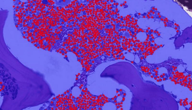
- Bone marrow cells
- Lesion-specific applications for liver, kidney, and brain

Aiforia® Studies
Cutting-edge AI-assisted image analysis capabilities supported by a study-centric GLP workflow
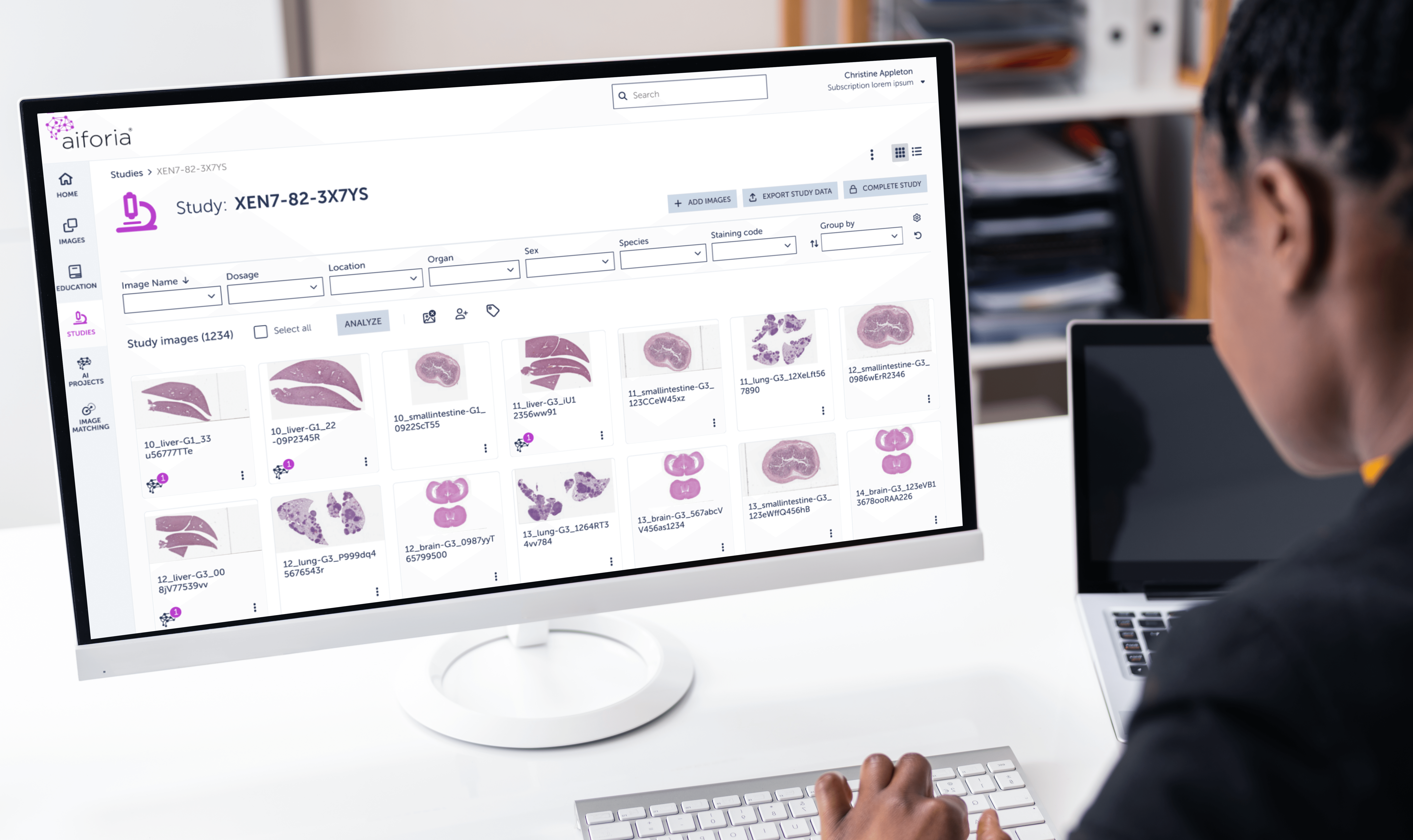
Aiforia® Studies is a software module that enables users to perform cutting-edge AI-assisted image analysis studies according to Good Laboratory Practices (GLP). Aiforia Studies is specifically designed for non-clinical studies that need to adhere to GLP guidelines, although its application is not restricted to such studies. The module enables users to perform and evaluate AI-assisted image analysis in non-clinical histology studies, and export and store immutable study data using the Aiforia® Platform.
Aiforia® Create
The power of AI in your hands
Aiforia® Create is the most versatile tool for developing, customizing, and validating deep learning AI models for histological features and patterns in image analysis.
Its cloud-based, collaborative working environment allows multiple users to work together in real time, anywhere in the world. Praised for its intuitive user interface, it allows users a fast start, even without any prior AI experience.
Comprehensive verification metrics are used to evaluate the performance of trained AI models, ensuring top quality.

Aiforia's research use cases

MIT case study: advancing lung cancer research with AI
Reseachers at the Tyler Jacks Lab, MIT, created artificial intelligence models to automate tumor grading as part of their lung cancer research studies.

Massachusetts General Hospital case study: AI-assisted image analysis of neurodegenerative disease markers
Researchers from Massachusetts General Hospital used Aiforia’s AI for the analysis of histopathological markers in neurodegenerative diseases.

Faron Pharmaceuticals case study: using AI to perform spatial analysis in cancer drug development
Elisa Vuorinen at Faron Pharmaceuticals built an AI model to quantify and localize Clever-1 in the tumor microenvironment using Aiforia® Create.

Sanofi case study: Parkinson's disease research with AI
The preclinical research team studying Parkinson's disease at Sanofi created their own AI model with Aiforia® Create to automate Th+ neuron quantification.

Orion pharma case study: accelerating preclinical neurotoxicity analysis with AI
Scientists at the pharmaceutical company Orion Pharma developed artificial intelligence models to automate preclinical neurotoxicity assessment.

CRL case study: AI-assisted screening of bone marrow cellularity changes
CRL Veterinary Pathologist Mark Smith describes using AI models to screen for bone marrow cellularity changes.

Experimentica case study: accelerating preclinical analysis of ocular diseases
Scientists at the CRO Experimentica describe using AI to analyze Spectral Domain Optical Coherence Tomography scans to identify neovascular lesions.
Our services
Integration services
Integration can be straightforward. Aiforia's solutions can be integrated with any existing laboratory infrastructure to enjoy the full benefits of a digitized workflow.
Cloud hosting services
Choose to host on our secure shared or private cloud environment. You can also opt to host on your own cloud provider (preferred partners include MS Azure, GCP, and AWS).
Custom AI services
AI models developed by scientists, for scientists. With custom AI services, Aiforia's scientists build AI models for your specific needs.
Request a demo
Discover the power of AI for image analysis
Find out how to enhance your image analysis work in diagnostic pathology, preclinical studies, and medical research. The demo will be tailored based on your interests.
The demo will help you understand:
- How AI-assisted image analysis can increase efficiency, precision, and consistency in the pathology workflow.
- The limitless possibilities Aiforia® Platform offers – both for research and clinical diagnostics – and the suitable use cases for your needs.
We can either demonstrate Aiforia’s image analysis solutions on your own images or any of our application examples (i.e., neuron quantification, automated tumor grading, NASH analysis, etc.).
Fill in the form, and one of our experts will contact you shortly to schedule the time.