Aiforia®️ Create is the most versatile tool for developing deep learning AI models for image analysis. This article explains its immunofluorescence imaging and analysis capabilities.
Unlimited channels, limitless data
Aiforia’s multichannel viewing and analysis capabilities can be applied to any image, allowing hundreds of channels to be imported and processed.
When viewing multichannel images, you can adjust the visibility (channel intensity) of each channel and select the colors you want to use to view the image. You can hide or highlight the channels of interest as you go.
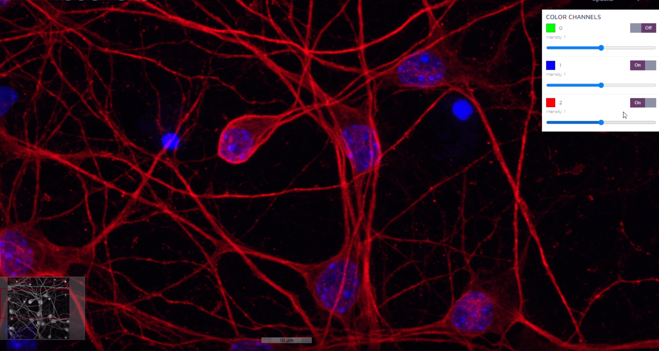
Multichannel immunofluorescent image of cultured neurons showing microtubule (red) and nuclear (blue) staining
The neural networks of our deep learning AI are able to learn from all channels at the same time. In particular, the neural networks are able to learn which channels are the most useful ones for a specific task. You can view and turn on/off as many channels as you want to increase feature visibility while annotating.
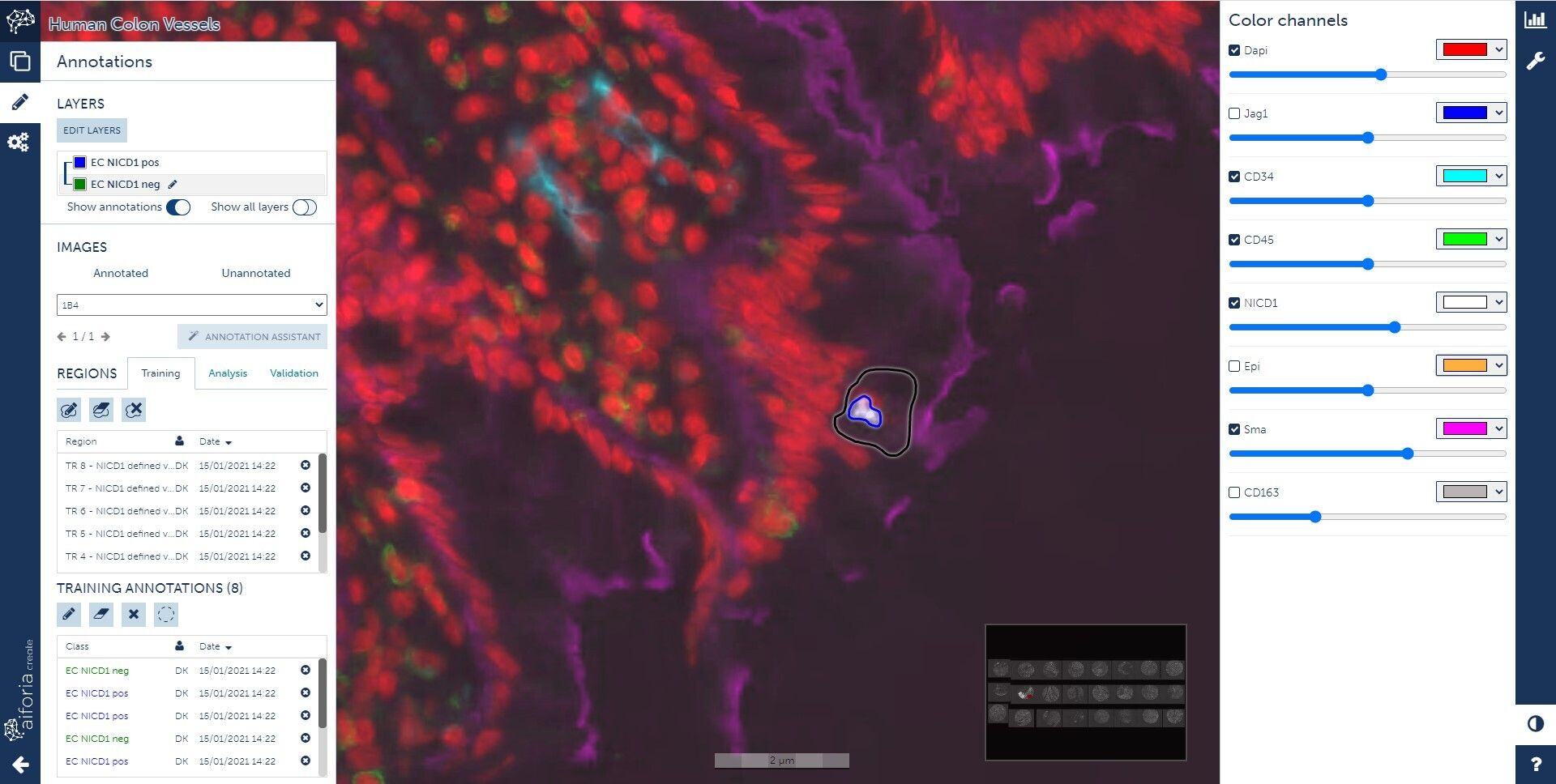
Aiforia Create's training window showing annotations for NICD1 in multi-channel (Immunofluorescence) human colon tissue, stained with various vascular & immune cell markers in addition to epithelium
Multichannel (IF) human colon tissue showing staining for various vascular and immune cell markers in addition to epithelium in Aiforia slide viewer:
Get to know other Aiforia Create features:
- Annotation Assistant
- Transfer learning
- Object detection, semantic segmentation, and instance segmentation
- Validation features
