• There are no suggestions because the search field is empty.

AI-powered veterinary diagnostics

Supporting pathologists working in veterinary diagnostic labs, the Aiforia® Platform offers an efficient case-centric workflow for viewing images and analyzing results. Aiforia’s AI applications assist decision-making and automate repetitive or complex tasks. Cases, findings, and markings can easily be shared with colleagues for consultation and collaboration.

Contact us
Aiforia_veterinary diagnostics-1


Unlock the full benefits of a digitized workflow

ic_speed_up_pink

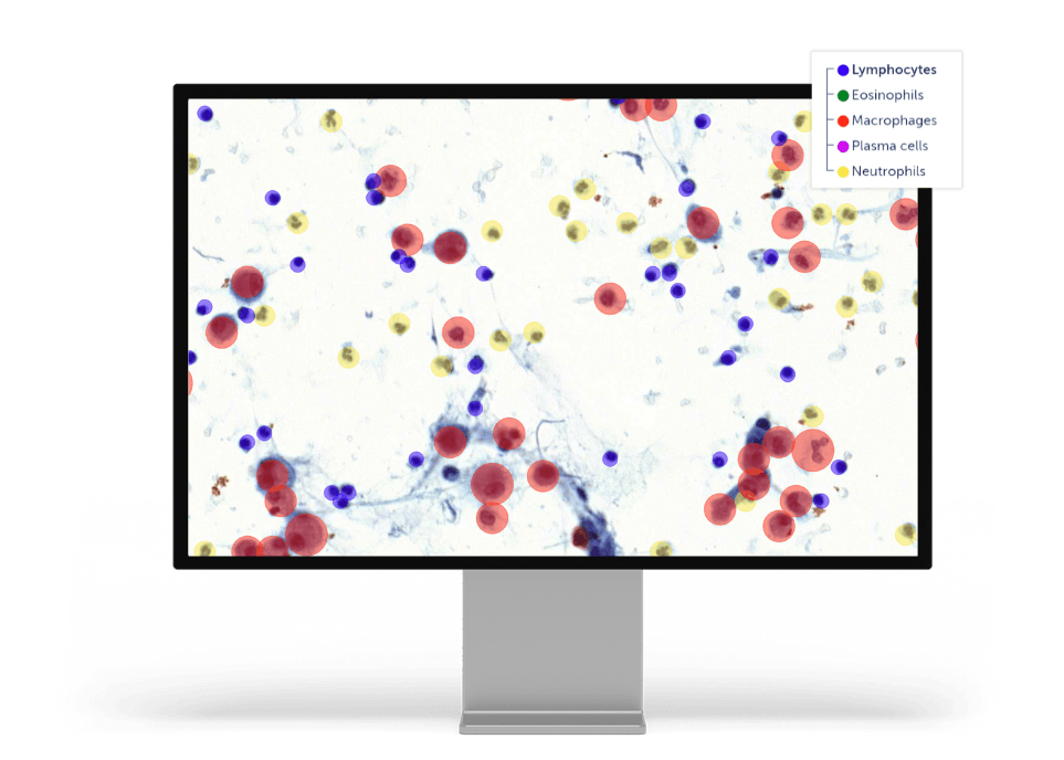
AI-supported quantification and scoring

Automation of repetitive or difficult tasks for a pathologist to review, adjust as needed, and sign out. 

ic_discover_pink

Efficient viewing of whole-slide images, hotspots and regions

Fast viewing of analysis results on whole slide images or selected areas for more precise examination and scoring. 

ic_our people-1_pink

Collaboration with colleagues

Share cases, findings, and markings easily for consultation and collaboration with colleagues. 

ic_our_ai-1_pink

Seamless integrations

Integrations with any existing laboratory infrastructure unlock the full benefits of a digitized workflow. 

Veterinary diagnostic AI applications_example 2

Veterinary diagnostic AI applications

Examples of applications to support veterinary pathology diagnostics 

  • Mitoses
  • Gastrointestinal
    • H. pylori in stomach samples
    • Immune cell counter
    • Goblet cell detector
  • Bronchoalveolar lavage cytology
  • Tumor margin
  • Tumor detector  
  • Bone marrow cell lineages

Aiforia® Create – AI development tool

Learn more about Aiforia's the most versatile tool for developing deep learning AI models for image analysis.

CASE EXAMPLE

Finn Pathologists case study: canine mast cell tumor – Ki-67 scoring

  • Mast cell tumor (MCT) is the most common skin tumor in dogs; it can also affect other areas of the body, including the spleen, liver, gastrointestinal tract, and bone marrow.
  • Ki-67 is a nuclear protein only expressed in cells that are dividing. Ki-67 expression can be detected with an antibody, measuring cell proliferation.
  • The frequency of Ki-67 expression is significantly associated with prognosis. 
  • Using outcome data, a cut-off threshold (Ki-67 labeling index) is calculated. Above the threshold, the prognosis for local recurrence and overall patient survival is poorer.


Veterinary Diagnostic AI Applications_Finn Pathologists_IHC - MIB-1 (Ki-67)_1_logo 1


Contact us

Discover the power of AI for pet diagnostics

Discover how Aiforia’s AI can enhance your veterinary diagnostic process, tailored to your lab’s unique needs.

We will help you understand:

  • How AI-assisted image analysis can increase efficiency, precision, and consistency in a case-centric workflow.
  • The limitless possibilities Aiforia® Platform offers and the suitable AI applications for your workflow.

Fill in the form, and one of our experts will contact you shortly.