Aiforia® Create – AI development tool
The power of AI in your hands

Using Aiforia® Create requires no data science or software programming expertise. Its cloud-based nature makes it scalable and extremely fast to implement; no installation is needed.
Aiforia® Create is compatible with any 2D image and a broad range of image file formats, including brightfield and fluorescence. It can be integrated with any existing laboratory infrastructure to enjoy the full benefits of a digitized workflow.
Thousands of AI models have been developed with Aiforia® Create for research and clinical use cases; applications ranging from cancer research to neuroscience and even outside the medical field.
Aiforia® Create supports the user by automating manual steps in AI development with features such as the patented Annotation Assistant.
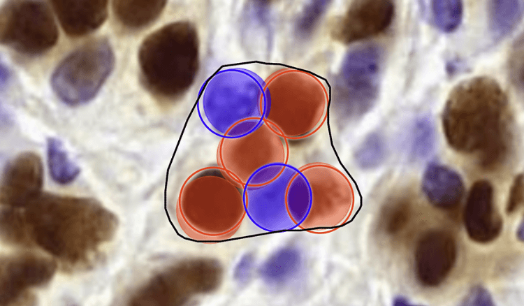
ANNOTATION ASSISTANT
The patented Annotation Assistant utilizes active learning, a highly sought-after technique in artificial intelligence. It offers premade annotations on the most useful areas of training data, allowing the user to accept, modify, or reject the suggestions.
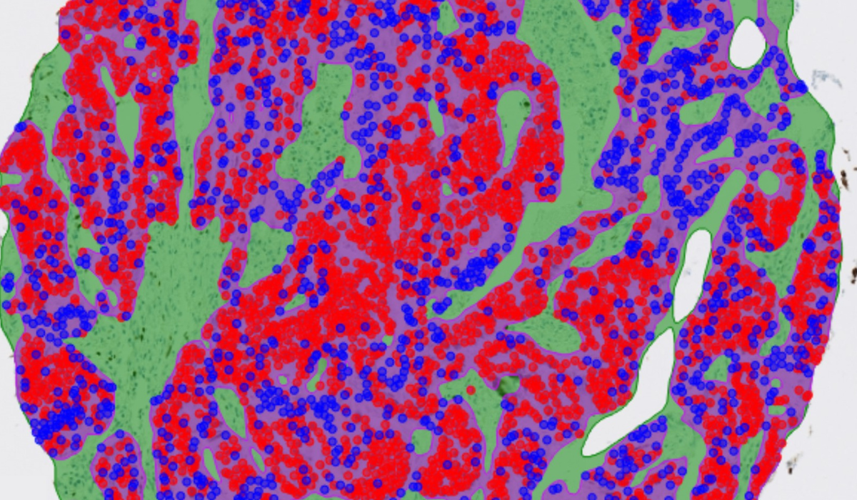
OBJECT DETECTION
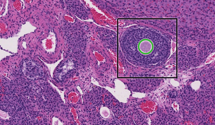
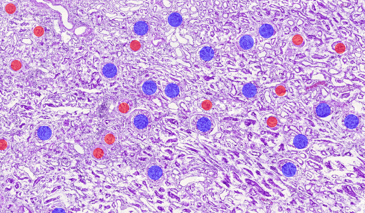
Aiforia’s object detection technology can detect thousands of different objects in complex images. It enables multi-class identification and the detection of rare objects and can be combined with segmentation and spatial metrics for enhanced analysis.
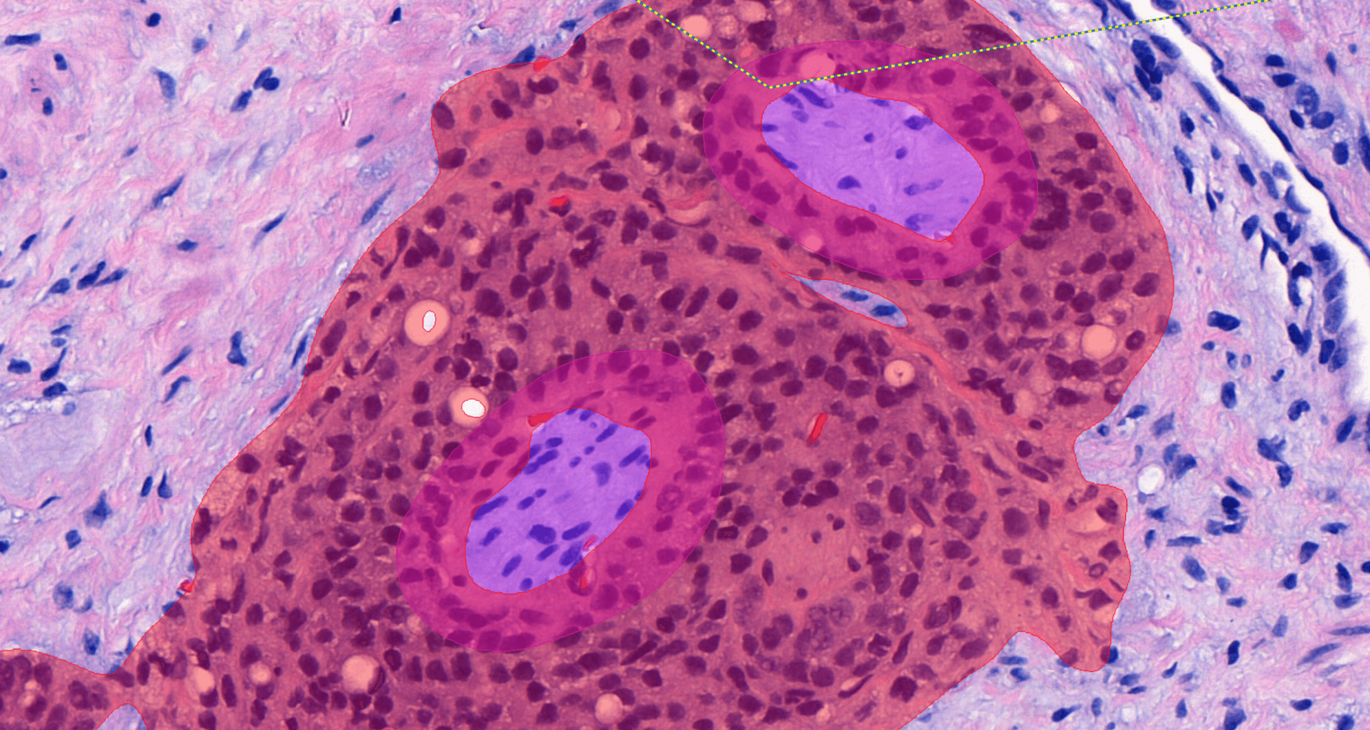
SEMANTIC AND INSTANCE SEGMENTATION
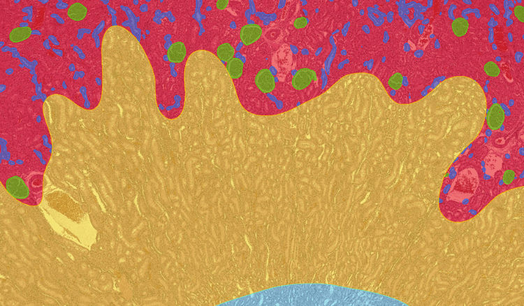
Segmentation features enable precise quantification of areas and shapes, even individually when areas merge or overlap. Analysis can be further boosted when combined with object detection and spatial metrics.
TRANSFER LEARNING
Transfer learning reduces the number of annotations and training cycles needed in AI development by allowing the use of existing models as a basis for AI model development and fine-tuning.
VALIDATION
Validation features provide an easy interface for defining validation sets and collecting validation annotations remotely. They also provide a convenient way to invite colleagues or consultants to give blinded scoring or diagnosis according to intended use criteria.
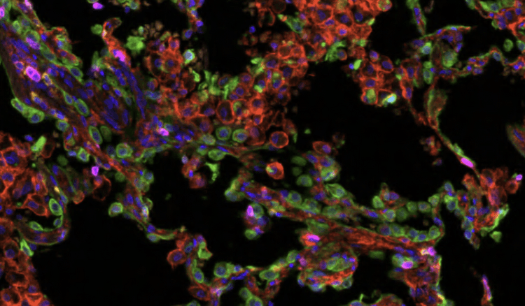
MULTICHANNEL IMAGES AND ANALYSIS
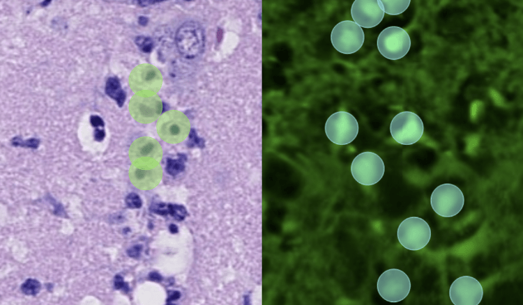
All AI development features are applicable to multichannel images. This enables the viewing and analysis of complex immunofluorescence images.

ANNOTATION ASSISTANT
The patented Annotation Assistant utilizes active learning, a highly sought-after technique in artificial intelligence. It offers premade annotations on the most useful areas of training data, allowing the user to accept, modify, or reject the suggestions.

OBJECT DETECTION
Aiforia’s object detection technology can detect thousands of different objects in complex images. It enables multi-class identification and the detection of rare objects and can be combined with segmentation and spatial metrics for enhanced analysis.

SEMANTIC AND INSTANCE SEGMENTATION
Segmentation features enable precise quantification of areas and shapes, even individually when areas merge or overlap. Analysis can be further boosted when combined with object detection and spatial metrics.

TRANSFER LEARNING
Transfer learning reduces the number of annotations and training cycles needed in AI development by allowing the use of existing models as a basis for AI model development and fine-tuning.

VALIDATION
Validation features provide an easy interface for defining validation sets and collecting validation annotations remotely. They also provide a convenient way to invite colleagues or consultants to give blinded scoring or diagnosis according to intended use criteria.

MULTICHANNEL IMAGES AND ANALYSIS
All AI development features are applicable to multichannel images. This enables the viewing and analysis of complex immunofluorescence images.
A research team from the University of Helsinki used Aiforia® Create to develop a deep learning AI model to predict patient outcomes in a complex ovarian cancer.

AI-assisted diagnosis was compared to visual diagnosis in a study involving H&E slides from 111 prostate cancer patients. Read about the results.
Dr. Jennifer M. Boland et al. from the Mayo Clinic successfully developed an AI model to determine the invasion in pulmonary adenocarcinoma.

Dr. Rish Pai from the Mayo Clinic developed an AI model to assist oncologists in deciding which CRC patients should receive chemotherapy and for how long.
/Cancer%20JPEG/colorectal-cancer_after.jpg)
Veterinary pathologists from the UK-based Finn Pathologists used Aiforia® Create in the Ki-67 scoring of canine mast cell tumors.

A research team from the University of Turin used Aiforia® Create to build an AI model for mesothelioma subtyping with reticulin stain. Read more or watch the video interview.

In this study, the results of Ki-67 scoring performed with Aiforia® Platform were compared against three independent pathologists on various solid tumors.
Reseachers at the Tyler Jacks Lab, MIT, created artificial intelligence models to automate tumor grading as part of their lung cancer research studies.
/Cancer%20JPEG/Tumor_grading_MIT_after.jpg)
A research team from Cerba Research compared the results of Ki-67 scoring performed with the Aiforia® Platform against two independent pathologists.

Pathologists can automate and improve the accuracy of calculating Ki-67 in any image. This case study describes the benefits of using the Aiforia software.

Liesbeth Hondelink, MSc student at Leiden University Medical Centre, describes using AI to automate the quantification of PD-L1 in lung cancer studies.
/Cancer/PD-L1%20before.png)
The preclinical research team studying Parkinson's disease at Sanofi created their own AI model with Aiforia® Create to automate Th+ neuron quantification.

PhD student Polina Stepanova discusses the benefits of using AI for mutant huntingtin detection to improve prediction methods and develop new therapies.

Researchers from Massachusetts General Hospital used Aiforia’s AI for the analysis of histopathological markers in neurodegenerative diseases.

Researchers at the University of Helsinki deployed artificial intelligence models to automate Parkinson’s disease neuron counting.

A neuroanatomy lab in Pamplona, Spain, uses Aiforia® Create to advance neurodegenerative disease research.

Interview with Duke University PhD Student on dopaminergic neuron quantification in Parkinson’s disease research.

Van Andel Institute created four AI models with Aiforia to provide unbiased, quantitative analysis for Parkinson’s disease research.
Parkinson’s disease, a neurodegenerative disorder, is commonly detected using brain scans and analyzed manually. We interviewed neuroscience researcher Joan on the benefits of AI in PD research.

Stereology has long been a reliable method for neuron quantification, but how does it compare in speed and accuracy to artificial intelligence based tools?

Dr. Maxwell L. Smith from Mayo Clinic built an AI model to accurately estimate large droplet fat in liver sections prior to transplantation.

This case study describes the use of AI in studying nonalcoholic fatty liver disease (NAFLD) and its capability to segment structures in liver histology.

A pathologist studying primary sclerosing cholangitis (PSC) created a deep learning AI model to evaluate novel prognostic biomarkers of the liver disease.

Dr. Rish Pai from the Mayo Clinic developed an AI model to assist oncologists in deciding which CRC patients should receive chemotherapy and for how long.
Dr. Maxwell L. Smith from Mayo Clinic developed two AI models to detect and quantify histopathologic features in interstitial lung disease (ILD) with the goal of using AI to help differentiate ILD subtypes.

Dr. Jennifer M. Boland et al. from the Mayo Clinic successfully developed an AI model to determine the invasion in pulmonary adenocarcinoma.
A research team from the University of Turin used Aiforia® Create to build an AI model for mesothelioma subtyping with reticulin stain. Read more or watch the video interview.
Reseachers at the Tyler Jacks Lab, MIT, created artificial intelligence models to automate tumor grading as part of their lung cancer research studies.
Kati Mäkelä, an MD specializing in pulmonary medicine, tells us about using AI to quantitate histopathological features of idiopathic pulmonary fibrosis.
/Other%20medical%20areas%20JPEG/lung_fibroblast_lymphocytes_after.jpg)
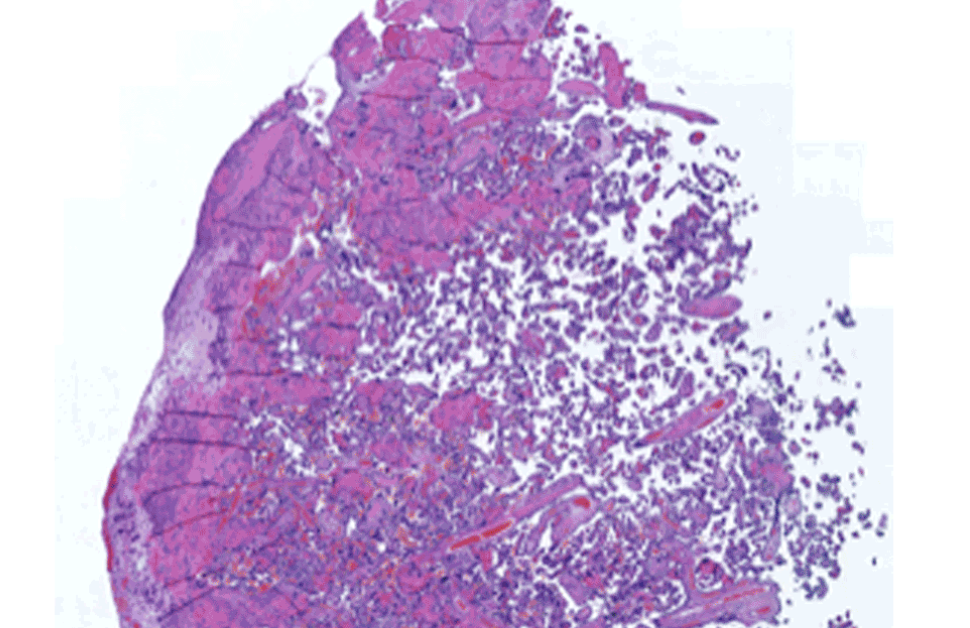
An Italian research team built an AI model for kidney pathology to assist on-call pathologists and renal pathologists in their routine work.
/Other%20medical%20areas%20JPEG/kidney_cortex_medulla_after.jpg)
A research team from the University of Helsinki used Aiforia® Create to develop a deep learning AI model to predict patient outcomes in a complex ovarian cancer.
Pathologist Dr. Leena Strauss developed an artificial intelligence model to evaluate the stages of the estrus cycle in cytology samples.
/Other%20medical%20areas/Reproductive%20disease.png)
Veterinary pathologist, Mark Smith, from Charles River Laboratories describes using AI models to screen for bone marrow cellularity changes.

Elisa Vuorinen at Faron Pharmaceuticals built an AI model to quantify and localize Clever-1 in the tumor microenvironment using Aiforia® Create.

Scientists at the pharmaceutical company Orion Pharma developed artificial intelligence models to automate preclinical neurotoxicity assessment.

Veterinary pathologist, Mark Smith, from Charles River Laboratories describes using AI models to screen for bone marrow cellularity changes.
Pathologists at Charles River Laboratories used Aiforia's AI development tool globally to automate preclinical assessments.
Scientists at the CRO Experimentica describe using AI to analyze Spectral Domain Optical Coherence Tomography scans to identify neovascular lesions.

View case studies of using deep learning AI in studying and diagnosing infectious diseases, such as malaria, tuberculosis, and hantavirus.

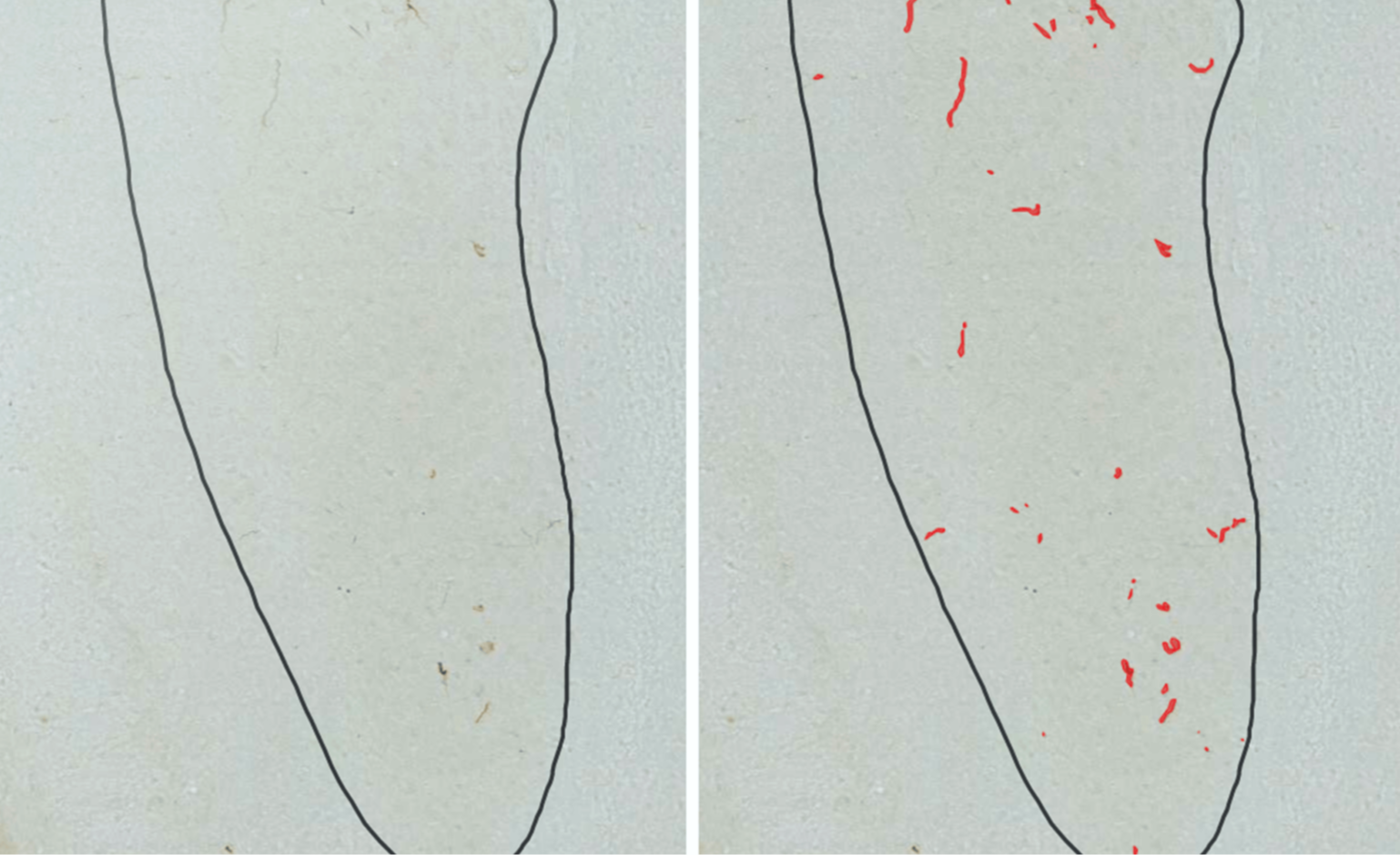
AI-assisted analysis of malaria parasites has the potential to significantly improve diagnosis and treatment of the deadly disease. Learn more.
/Other%20medical%20areas/Malaria%20after.png)
Interview with virologist Tomas Strandin on the use of AI in the quantification of antibodies signaling orthohantavirus infections

/Cancer/PD-L1%20after.png)
/Neuro%20JPEG/iron_detection_after.jpg)
/Neuro%20JPEG/astrocytes_orion_before.jpg)
/Liver%20JPEG/PSC_before.jpg)
/Other%20medical%20areas/DSS-colitis-2-after-1.png)
/Other%20medical%20areas%20JPEG/tuberculosis_after.jpg)
/Other%20medical%20areas/Reproductive%20endometrial%20after.png)
/Other%20medical%20areas%20JPEG/malaria_RBC_after.jpg)
/Other%20medical%20areas/Coronavirus%20after.png)
/Other%20medical%20areas/Tuberculosis%20after.png)
/Other%20industries/Salmon%20skin%20after-1.png)